Lab tests
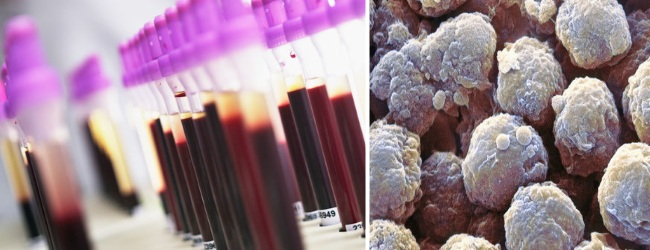
Alanine Aminotransferase
Enzyme ALT - high enzyme activity observed in skeletal muscle, heart, kidney and especially the liver. Determination of enzyme activity in the blood is often used for diagnostic and therapeutic uses for the assessment of liver cells - hepatocytes.
Normal figures: men - 21-72 U / l (U / L)
Women - 9-52 U / l (U / L)
Increased activity: rapid increases in necrosis of hepatocytes, hepatitis, liver tumors and metastases in the liver, liver cirrhosis, infectious mononucleosis, extensive skeletal muscle injury, myositis, myocarditis, and myocardial infarction, when taking certain medicines.
Lowering Activity: significant diagnostic value has not.
Preparation for the study: special training is required. To study procedures performed taking venous blood.
Research Method: colorimetric kinetic method using a dry multilayer analytical element analyzer gated VITROS 5.1, USA.
Aspartate Aminotransferase
The enzyme AST - high activity observed in heart, skeletal muscle and liver. Most often, the enzyme activity determination conducted in the diagnostic and therapeutic uses for the assessment of liver cells and heart muscle.
Normal figures: men - 17-59 U / l (U / L)
female - 14-36 U / l (U / L)
Increased activity: myocardial infarction, pulmonary embolism, injuries of muscles, cirrhosis, hepatitis, liver tumors.
Lowering Activity: significant diagnostic value has not.
Preparation for the study: special training is required. To study procedures performed taking venous blood.
Research Method: colorimetric kinetic method using a dry multilayer analytical element analyzer gated VITROS 5.1, USA.
Amylase
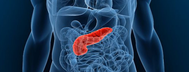
Amylase - the digestive enzyme responsible for the breakdown of carbohydrates food. It is produced by the pancreas and salivary glands. Due to the difficulty of diagnosis of acute pancreatitis laboratory determination of amylase activity in blood and urine has an important and sometimes crucial for diagnosis.
Normal performance: adult blood - 30-110 U / l (U / L)
urine - 32-641 U / l (U / L)
Increased activity: acute pancreatitis, abscess, trauma, tumors of the pancreas, mumps ( "pig") and renal failure.
Lowering Activity: serum and urine may be pancreatic necrosis.
Preparation for the study: special training is required. For the procedure, studies carried out taking venous blood and (or) spontaneous urine sample.
Research Method: colorimetric kinetic method using a dry multilayer analytical element analyzer gated VITROS 5.1, USA.
Albumin
Albumin - basic protein in the human body, produced in the liver. It contributes to the maintenance of osmotic pressure (water retention in the bloodstream) and is involved in the transportation of many compounds. It is believed the first biochemical marker of eating disorder.
Normal figures: 35-50 g / l.
Increase in concentration: almost does not occur, may indicate dehydration.
concentration Lowering: insufficient intake of protein with food (starvation, malnutrition), impaired protein absorption in the gastrointestinal tract (bowel disease, surgical removal of part of the intestine or the stomach), decreased albumin synthesis in the liver (chronic hepatitis, cirrhosis, liver tumor) increased protein loss (ulcerative colitis, peritonitis, extensive burns), kidney damage (nephrotic syndrome with the presence of protein in the urine). albumin content is usually reduced in chronic inflammatory diseases, pregnancy.
Preparation for the study: special training is required. To study procedures performed taking venous blood.
Research Method: colorimetric method using a dry multilayer analytical element analyzer gated VITROS 5.1, USA.
Alpha-fetoprotein (AFP)
Alpha-fetoprotein, AFP, alpha-fetoprotein (fetal alpha globulin, AFP) - one of the first tested and proven tumor markers. AFP also applies vprenatalnoy diagnosis.
Normal performance: up to 8 IU / ml.
Increased blood levels of AFP with tumor diseases:
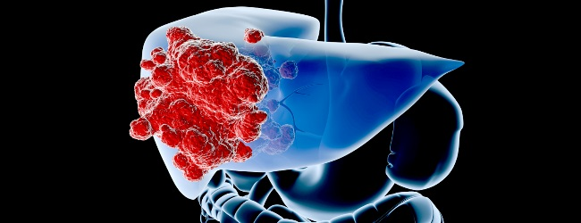
Primary liver cancer - hepatoblastoma or gepatotselyullyarny cancer;
tumors of the germinal cells of the ovaries and testes - must be controlled simultaneously with hCG.
- Increasing the level of AFP under these tumors has a high sensitivity and specificity, which makes analysis of AFP in cases of suspected disease data MANDATORY!
- tumor metastases to the liver;
bronchogenic carcinomas;
mammary cancer;
gastric cancer (AFP analysis is carried out simultaneously with the CA 72-4);
colon cancer;
- pancreas cancer.
AFP also increases in other diseases, noncancerous etiology:
cirrhosis of the liver;
acute viral hepatitis;
chronic hepatitis;
chronic renal failure.
A transient (temporary) increase in the rate of AFP in the blood in benign diseases:
steatosis;
liver cysts;
adenoma of the liver;
nodular hyperplasia of the liver;
cholecystitis - inflammation of the gallbladder;
cholelithiasis;
active liver regeneration (e.g., after administration of antibiotics and antivirals).
Indications for a blood test to AFP:
diagnosis and monitoring the effectiveness of the treatment of primary liver cancer (hepatoblastoma and hepatocellular carcinoma);
diagnosis and monitoring the effectiveness of treatment of patients with tumors of germ cell origin (including hCG);
AFP analysis carried out in the course of examination of patients at high risk of liver cancer - in people with a positive test for HBs-Ag and liver cirrhosis for early detection of malignancy;
identification of fetal malformations - neural tube defects and abdominal wall, Down's syndrome.
Features a blood test to AFP:
physician recommendation is to be tested for AFP - a necessary precaution;
research should be done in the dynamics - in the process of diagnosis, before and after treatment;
obligatory simultaneous analysis of other oncomarkers
analysis should be carried out in the same laboratory, by the same method;
for the correct decryption result of the analysis must take into account in the AFP data from other research methods (CT, ultrasound, biopsy);
raising the level of AFP in the blood - NOT A diagnosis of cancer!
Tumor markers CEA (carcinoembryonic antigen, CEA)
Tumor markers CEA -glycoprotein high molecular weight.Determined in elevated concentrations in 60% of patients with colon cancer.
Normal figures: 0-5 ng / ml.
On the level of CEA affected by smoking and to a lesser extent alcohol intake (increase the level of the marker).
Smoking and CEA
97% non-smoking healthy plasma CEA concentrations less than 3.0 ng / ml;
90% of smokers and ex-smokers 7% in the plasma concentration of CEA than 3.0 ng / ml.
application
Monitoring of persistent, recurrent or metastatic colonic adenocarcinoma; is elevated in more than 30% of patients with adenocarcinoma of the breast, lung, liver cancer, carcinoma of the pancreas.
Performance indicator surgery, chemotherapy and radiation therapy
Prediction for patients with colon cancer.
Not recommended for screening due to low sensitivity / specificity, especially in the early stages of malignancy, as carcinoembryonic angiten reflects a significant amount of the tumor mass.
Diagnosis of malignant pleural effusion.
CEA also increased in other diseases noncancer etiology:
liver diseases (hepatitis);
pancreatitis;inflammatory bowel disease (ulcerative colitis, Crohn's disease);
chronic lung disease;
autoimmune disease.
Features a blood test to CEA
physician recommendation is to be tested for CEA - a necessary precaution;
research should be done in the dynamics - in the process of diagnosis, before and after treatment;
obligatory simultaneous analysis of other oncomarkers;
analysis should be carried out in the same laboratory, by the same method;
for the correct decryption result of the analysis at the CEA must take into account the findings of other research methods (CT, ultrasound, biopsy);
raising the level of CEA in the blood - NOT A diagnosis of cancer!
Research: enhanced chemiluminescence analyzer gated VITROS ECiQ, USA.
Tumor markers CA 19-9
Tumor marker CA 19-9 - tumor associated antigen whose level most strongly increased in patients with pancreatic cancer and certain tumors of the gastrointestinal tract and ovary. At 10.7% of people in the population (absence of a particular gene) is not synthesized even in the presence of the tumor.
Normal performance: up to 37 IU / mL.
Increased CA 19-9 level with tumor diseases
adenocarcinoma of the pancreas;
gastric tumor;
carcinoma of the colon;
biliary tract neoplasms;
swelling of the liver - holangiotselyullyarnaya carcinoma;
certain tumors of the female reproductive system, often with mucosal tumors of the ovary.
CA 19-9 is also increased in other diseases, noncancerous etiology:
pancreatitis,
hepatitis,
cirrhosis of the liver,
endometriosis,
uterine fibroids.
Indications for analysis on CA 19-9
Diagnosis of the disease - due to the high sensitivity is used primarily for the early diagnosis of pancreatic tumors;
assessment of the severity of the disease - tumor volume does not correlate with the concentration of CA 19-9, but the rapid growth of tumor marker above 10 000 IU / L
indicates a metastasis;
monitoring treatment success.
Features a blood test SA19-9
physician recommendation is to be tested for CA 19-9 - a necessary precaution;
research should be done in the dynamics - in the process of diagnosis, before and after treatment;
obligatory simultaneous analysis of other oncomarkers;
analysis should be carried out in the same laboratory, by the same method;
to properly decrypt the result of analysis on the CA 19-9 is necessary to take into account the findings of other research methods (CT, ultrasound, biopsy);
improvement of CA 19-9 in the blood - NOT A diagnosis of cancer!
Research: enhanced chemiluminescence analyzer gated VITROS ECiQ, USA.
Tumor markers CA 125II
Cancer antigen CA 125 glycoprotein synthesized by the serous membrane of internal body cavities, and some bodies. In women of childbearing age the main source of the SA-125 is the endometrium, what has caused the cyclical variation in blood tumor marker depending on the phase of the menstrual cycle. CA-125 concentration is increased in the menstruation period, in the first trimester of pregnancy. Elevated levels of CA -125 is found in approximately 80% of women with ovarian cancer.
Normal performance: up to 35 IU / mL.
Indications for the analysis of CA 125:
Identification of an epithelial ovarian cancer.
All women with a history of a history of ovarian cancer - a close relative suffered from ovarian cancer.
Monitoring the success of the treatment. In most cases, the beginning of raising the CA 125 blood ahead of the clinical manifestation of relapse of disease for 4-6 months.
Detection of metastases.
Analysis of CA 125 is not used for screening for ovarian cancer.
Increasing the level of CA 125 with tumor diseases:
ovarian cancer;
endometrial cancer;
cervical cancer;
breast cancer;
lung cancer;
pancreas cancer;
tumors of the gastrointestinal tract;
metastases to these organs.
CA 125 is also increased in other diseases noncancer etiology:
peritonitis,
pleurisy,
pericarditis,
ovarian cysts,
benign hyperplasia of the endometrium.
Reduced levels of CA125
reduced level of CA125 - rate;
tumor marker in the blood has not yet risen to the diagnostically significant parameters;
the tumor tissue does not synthesize CA 125;
the success of the treatment.
A negative test result in the CA 125 is not evidence of absence of cancer.
Features a blood test CA-125
physician recommendation is to pass on the analysis of CA 125 - a necessary precaution;
research should be done in the dynamics - in the process of diagnosis, before and after treatment;
obligatory simultaneous analysis of other oncomarkers;
analysis should be carried out in the same laboratory, by the same method;
to properly decrypt the result of analysis on the SA 125 is necessary to take into account the findings of other research methods (CT, ultrasound, biopsy);
increase in CA-125 blood - NOT A diagnosis of cancer!
Research: enhanced chemiluminescence analyzer gated VITROS ECiQ, USA.
Tumor markers CA - 15-3
The primary mammary tumor marker. Increasing the concentration indicates the presence of tumors, but do not exclude the negative pathology. Required study in dynamics, disposable test result is not informative.
Normal performance: up to 35 IU / mL.
Indications for the analysis of CA 15-3
assessment of the severity of the disease - tumor volume correlated with the concentration of CA 15-3,, the rapid growth of tumor marker indicates metastasis;
monitoring during treatment - response to treatment ,;
control of the total treatment success upon its completion;early diagnosis of recurrence;
Diagnosis of the disease - due to the high sensitivity is used primarily for the early diagnosis of breast tumors when indicated.
Analysis of CA 15-3 is not used as a screening - not all women examined without reading!
Increased CA 15-3 level with tumor diseases
breast cancer - carcinoma, adenocarcinoma;
lung cancer, bronchogenic carcinoma;
tumors of the gastrointestinal tract;
livercancer -gepatotsellyulyarnayacarcinoma;
pancreascancer;
prostatetumors;
ovariantumors, uterinecancer, cervicalcancer.
Increased CA 15-3 level with non-neoplastic diseases
benign tumors in the breast - 20% of cases (breast, fibroadenoma);liver disease - cirrhosis of the liver, acute viral hepatitis, chronic viral hepatitis, an inflammation of the biliary tract (cholangitis);
lung disease - chronic bronchitis, bronchial asthma, pneumonia;
kidney disease - chronic renal failure;
rheumatic diseases.
Reduced levels of CA15-3
the absence of the disease - the norm;
early stage breast cancer, when the tumor marker in the blood has not yet risen to the diagnostically significant parameters;
the success of the treatment.
A negative test result CA15-3 is not proof of absence of cancer.
Features CA15-3 blood test physician recommendation is to be tested for CA 15-3 - a necessary precaution;
research should be done in the dynamics - in the process of diagnosis, before and after treatment;
obligatory simultaneous analysis of other oncomarkers;
analysis should be carried out in the same laboratory, by the same method;
to properly decrypt the result of analysis on the CA 15-3 is necessary to take into account the findings of other research methods (CT, ultrasound, biopsy);
improvement of CA 15-3 in the blood - NOT A diagnosis of cancer!
Research: enhanced chemiluminescence analyzer gated VITROS ECiQ, USA.
total PSA
Prostate-specific antigen, or PSA - prostate primary tumor marker, in medical practice used for early diagnosis of prostate cancer. It is specific to the prostate gland.
Rates of PSA in the blood of men
0-50 years - to 2.5 ng / ml;
50-60 - 3.5 ng / ml;
60-70 years - to 4.5 ng / ml;
70-110 years - do6,5 ng / mL.
PSA index in the range of 4-10 ng / ml - the so-called "gray zone" - can be a sign of disease and physiological norm. For a more accurate assessment of applied research of free PSA and its ratio to the total. Normally, the amount of free PSA is not more than 20-25% of the total.
The practice is also used PSA growth rate for the year. In a normal year increase in PSA should not exceed 0.75 ng / mL. Large numbers are an indication for biopsy.
Indications for analyzing PSA
all men aged 40, 45, and 50 years after the PSA test should be performed 1 time per year;
if a family history of prostate cancer a man has to be tested for PSA 1 per year after 40 years;
the detection of prostate enlargement or other changes in her rectal examination;
men with symptoms of prostate cancer - pain and frequent urination, weak stream of urine, burning and discomfort during and after urination, pain in the bones of the pelvis;
to assess the severity of prostate cancer;
the success of the treatment and control of metastatic prostate cancer.
Increased PSA levels during tumor diseases
Prostate cancer - the underlying disease in which blood PSA level increased to a diagnostically significant parameters.
the PSA test should be evaluated together with the results of digital examination of the prostate and ultrasound. The definitive diagnosis of prostate cancer doctor will be able to put only on the basis of a biopsy - taking a small piece of prostate tissue and its study under a microscope.
PSA is not directly diagnose prostate cancer. "The gold standard" diagnosis of prostate carcinoma - biopsy. However, the indication for prostate biopsy is to increase the PSA level in the blood!
Features of the analysis of PSA
exercise and manipulation of the prostate increases PSA levels in the blood - finger issleddovanie prostate, bladder catheterization, cycling, sex - only 0.2 ng / ml, which has no diagnostic value;
vitamin H (biotin) at a dose higher than 5 mg / day - need to conduct analysis after 8 hours after administration of the tablet;
the amount of tumor tissue in direct proportion to the level of PSA in the blood.
Increased PSA with non-neoplastic diseases
BPH - benign diseases in which increased prostate size and there are problems with urination, more often in men older than 50 years;
inflammation of the prostate - prostatitis (both acute and chronic);
myocardial ischemia or prostate.
Research: enhanced chemiluminescence analyzer gated VITROS ECiQ, USA.
Bilirubin total
Bilirubin total - the total amount of indirect (unconjugated), direct (conjugated) bilirubin and delta bilirubin bound to blood proteins ..The basis of the formation of bilirubin - the disintegration of hemoglobin and other heme-containing proteins and enzymes. Since the increase in total bilirubin fractions observed in various pathological conditions, to clarify the causes of increased total bilirubin must also undertake to determine the concentration of direct and indirect bilirubin.
Normal indicators: adults - 3-22 mmol / l.
Increasing the concentration of: liver cell damage (hepatitis, cirrhosis, cancer), violation of the outflow of bile (calculous koletsistit, obstruction of the intra- and extrahepatic bile ducts, pancreas tumor), hematological diseases, congenital disorders of bilirubin metabolism, neonatal jaundice, taking certain medications .
Lowering Activity: diagnostic value has not.
Preparation for the study: special training is required. To study procedures performed taking venous blood.
Test method: colorimetric method using a dry multilayer analytical element gated analyzer VITROS 5.1, USA.
Bilirubin fractions
The fractions of bilirubin - conjugated and unconjugated bilirubin. Unconjugated bilirubin is formed by the decay of erythrocytes as a result of the biochemical reactions. Once the bloodstream to the liver, unconjugated bilirubin enters liver cells - hepatocytes, where it undergoes conjugation. The resulting conjugated bilirubin secreted into bile with it enters into the intestine and excreted.
Normal performance: adult unconjugated bilirubin - 0-19 mmol / l
conjugated bilirubin - 0-5mkmol / l
Increasing concentrations of unconjugated bilirubin: enhanced disintegration of red blood cells (haematological diseases fiziologicheskaaya jaundice neonatal jaundice of prematurity), liver cell damage (hepatitis, cirrhosis, cancer)
Increasing the concentration of conjugated bilirubin: violation of the outflow of bile (calculous koletsistit, obstruction of the intra- and extrahepatic bile ducts, pancreas tumor), liver cell damage (hepatitis, cirrhosis, cancer).
Lowering Activity: diagnostic value has not.
Preparation for the study: special training is required. To study procedures performed taking venous blood.
neonatal bilirubin
Bilirubin Neonatal - the sum of conjugated and unconjugated bilirubin. The test used for the detection of hyperbilirubinemia in newborns.
Normal figures: newborn 17-180 mmol / l.
Increase in concentration: neonatal jaundice.
Lowering Activity: diagnostic value has not.
Preparation for the study: special training is required. To study procedures performed taking venous blood.
Research Method: colorimetric kinetic method using a dry multilayer analytical element analyzer gated VITROS 5.1, USA.
Test method: colorimetric method using a dry multilayer analytical element gated analyzer VITROS 5.1, USA.

