Mammography
Mammography is a specific type of breast imaging that uses low-dose x-rays to detect cancer early – before women experience symptoms – when it is most treatable.
Your doctor would recommend a screening mammogram to detect breast cancer before it can be felt as a lump.
The aim is to detect breast cancer at its earliest stage. Early detection increases the likelihood of a cancer being successfully treated and often allows for greater treatment options. Screening mammography has been shown to decrease the death rate from breast cancer.
No preparation is required for screening mammography.
When you make your appointment, please let the radiology facility know if you have breast implants, so they can schedule a longer appointment. This is because it takes more time to obtain clear
X-ray images or pictures when implants are present.
Do not wear any deodorant, perfume, lotion or talcum powder on the day of your appointment, because these substances might show up as shadows on your mammogram. Wear a two-piece outfit, so you only need to undress from the waist up.
How is the procedure performed?
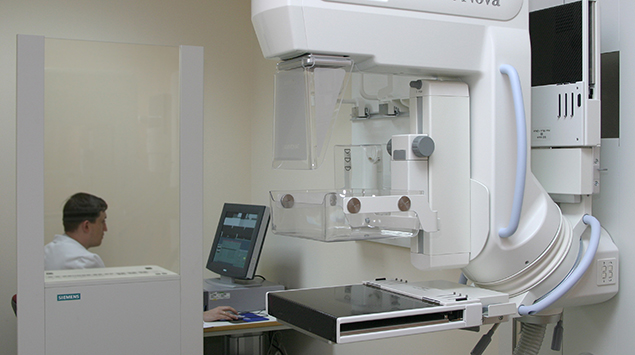
Mammography is performed on an outpatient basis.
During mammography, a specially qualified radiologic technologist will position your breast in the mammography unit. Your breast will be placed on a special platform and compressed with a clear plastic paddle. The technologist will gradually compress your breast.
Breast compression is necessary in order to:
-
Even out the breast thickness so that all of the tissue can be visualized.
-
Spread out the tissue so that small abnormalities are less likely to be hidden by overlying breast tissue.
-
Allow the use of a lower x-ray dose since a thinner amount of breast tissue is being imaged.
-
Hold the breast still in order to minimize blurring of the image caused by motion.
-
Reduce x-ray scatter to increase sharpness of picture.
You will be asked to change positions between images. The routine views are a top-to-bottom view and an angled side view. The process will be repeated for the other breast. Compression is still necessary for tomosynthesis imaging in order to minimize motion, which degrades the images. During screening breast tomosynthesis, two-dimensional images are also obtained or created from the synthesized 3-D images.
You must hold very still and may be asked to keep from breathing for a few seconds while the x-ray picture is taken to reduce the possibility of a blurred image. The technologist will walk behind a wall or into the next room to activate the x-ray machine.
When the examination is complete, you may be asked to wait until the radiologist determines that all the necessary images have been obtained.
The examination process should take about 30 minutes.
What will I experience during and after the procedure?
You will feel pressure on your breast as it is squeezed by the compression paddle. Some women with sensitive breasts may experience discomfort. If this is the case, schedule the procedure when your breasts are least tender. Be sure to inform the technologist if pain occurs as compression is increased. If discomfort is significant, less compression will be used. Always remember compression allows better quality mammograms.

